Today's Medical Mysterie
-
https://pubs.rsna.org/doi/10.1148/radiol.230033
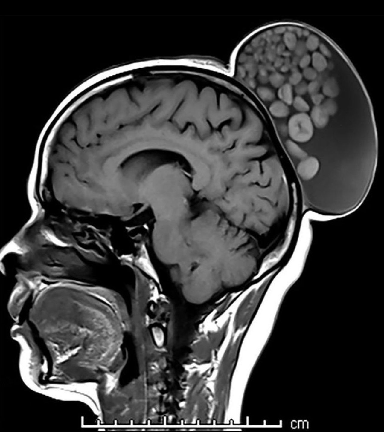
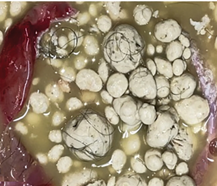
A 52-year-old woman presented with a painless, gradually enlarging scalp swelling that had been present since childhood. At examination, there was a 15 × 10 × 12–cm mass in the parieto-occipital region. MRI scan showed a large cystic lesion in the subgaleal plane of the scalp. It was hypointense with T1-weighted images and hyperintense with T2-weighted images and contained multiple nodules of varying sizes floating in the cyst, giving it a “sack of marbles” appearance (Figure). The nodules had thick outer rims that were iso- to hyperintense with T1-weighted sequences and hypointense with T2-weighted sequences and thin inner cores that were hypointense with T1-weighted sequences and hyperintense with T2-weighted sequences. The nodules demonstrated restricted diffusion (Figure) and had low apparent diffusion coefficient values. After completing surgical excision, the cyst was found to contain sebum-like material, hard spherules, and multiple strands of hair,
***Gross Medical Stuff***
click to show
Hello! It looks like you're interested in this conversation, but you don't have an account yet.
Getting fed up of having to scroll through the same posts each visit? When you register for an account, you'll always come back to exactly where you were before, and choose to be notified of new replies (either via email, or push notification). You'll also be able to save bookmarks and upvote posts to show your appreciation to other community members.
With your input, this post could be even better 💗
Register Login